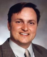
 Edward F. Fogarty III, MD, also known as “Ted” is a practicing clinical Diagnostic Radiologist with a procedural component in image-guided Interventional/Pain Management who practices primarily in North Dakota. He is the past Chairman of the Department of Radiology at the University of North Dakota (UND) School of Medicine and remains on faculty volunteering his time to continue in teaching residents and students affiliated with the UND Southwest Campus in Bismarck as well as the UND Northeast Campus in Grand Forks.
Dr. Fogarty has one of the more diverse practices of medicine via radiology and hyperbaric mitochondrial resuscitation techniques in all of the United States and Canada. He is an imaging research specialist who was part of the team that published the LSU Phase 1 Safety Trial for the use of Hyperbaric Oxygen Therapy (HBOT) in Post Traumatic Stress Disorder/ Traumatic Brain Injury (PTSD/TBI). He has several paradigm-shifting case reports involving medical imaging documentation of hyperbaric brain recovery protocols in conjunction with Paul G. Harch, MD at Louisiana State University in New Orleans who is one of two primary mentors in mitochondrial medicine.
Dr. Fogarty’s first publication in the medical literature was with a team assembled at Creighton University in Omaha, Nebraska reporting the spontaneous resolution of a large arteriovenous malformation discovered in utero on routine fetal ultrasound screening during the second trimester. The case presented during his time as Chief Resident at Creighton, while his mentor in Neuroradiology, Matthew Omojola, MBBS was Vice Chairman of the Department of Radiology at Creighton. This first experience with anomalous findings in medical data sparked a passion for documenting and deriving important scientific understanding out of “Black Swan” cases within information streams in medicine.
The art of teasing out the informative anomaly is an incredibly powerful point in science and medicine. Penicillin of course was discovered by just such an observation of an anomalous data point. Dr. Fogarty’s role in medicine is figuratively that of a photojournalist capturing black swans in research and his “day job” is in documenting all the rest of the flock on the radar screens in establishment academic medicine.
As fate would have it, this “bird dog” of imaging was identified in 2007 via a small network of political operatives and engineers with ties from Washington DC to North Dakota and Oklahoma and New Orleans, Louisiana (NOLA) affiliated with the International Hyperbaric Medical Foundation. The IHMF team connected Dr. Fogarty with Dr. Harch in New Orleans while Dr. Fogarty was visiting his brother, Robert X. Fogarty. This seminal meeting for the academic leadership of the University of North Dakota (UND) and Louisiana State University (LSU) in Hyperbaric Oxygen Therapy (HBOT) serendipitously occurred during the development of humanitarian hurricane disaster transportation services (evacuteer.org) by Dr. Fogarty’s brother, Robert in NOLA in conjunction with state and municipal political leadership.
While Dr. Fogarty was at the LSU-NOLA medical campus and at Dr. Harch’s clinic for 3 days, he was brought in to decode and understand the data and statistical deviations in Dr. Harch’s two-decade long accumulation of single-photon emission computed tomography (SPECT) data on multiple patients undergoing HBOT. Dr. Harch had already presented some of this data to the Department of Defense and Congress, several times, in efforts to generate funding for research in traumatic brain injured veterans in the years prior to their meeting. However, no one understood the significance of Dr. Harch’s breakthrough treatment.
When Dr. Fogarty saw the image data on local computer workstations in NOLA, he recognized that Dr. Harch had made a seminal discovery in his SPECT data from the very first time that these neurofunctional images started to be used in the 1980s. At that time, no one in academic radiology could translate those observations by Dr. Harch from film copy to digital imaging statistically analytics in the way Fogarty ultimately developed with Dr. Harch from 2007 to 2019. It was Ted’s task to statistically render what Dr. Harch had described to him as “smoothing” of the data.
The art of the practice of neurological hyperbaric medicine, which emerged during the 1980s in America, has accumulated documentation of incredible reversals of strokes and severely injured lobes of the brain. These are documented by SPECT images. These large area changes were obvious to all in medicine but what Dr. Harch was astutely observing was a more granular shift in all the data points rendered in these HMPAO-SPECT images.
Because of Dr. Ted Fogarty’s understanding of pioneering imaging informatics on a technical level, he quickly encoded the “smoothing” effect as a simple normalization on a bell curve of data. Less deviation from the mean in any area relative to an adjacent area is quite simply a reflection of this smoothness versus highly granular “pixelated” data. This granularity is a common phenomenon we all can see in under-exposed images from digital photography.
Dr. Fogarty understood what Dr. Harch was seeing; namely, he recognized a similar effect that the Impressionists such as Monet had obtained by using similar granulation/textural techniques. He was, therefore, able to re-encode the statistical rendering of granular data presentation in a way that others in medicine might more easily grasp. Dr. Ted Fogarty understood these concepts in the “gestalt” from homeschooling in Art/Art History by former Creighton University faculty member Mary Beth Schmidt Fogarty, his mother.
Many of the assembled scientific and medical luminaries on the advisory board have helped inform Dr. Fogarty’s practice immensely, learning through Hippocratic practice and exchange of ideas. This is how medicine has advanced over centuries. Dr. Fogarty has been in a close academic and advisory role with a luminary psychiatrist of New York over the last 4 years named Albert B. Crum, MD. Dr. Fogarty’s functional imaging career is a derivative of mitochondrial imaging. Mitochondrial Medicine is an emerging field of the 21st Century; Dr. Crum has mentored him into understanding the critical role of mitochondria and glutathione in detoxification as well as neurological regeneration via HBOT techniques.
Drs. Crum and Fogarty are currently working on pioneering strategies to accelerate metals detoxification in the simplest and safest of ways based on the development of oxygen in earth’s atmosphere and the genomic response to this event from billions of years past, and the toxic impacts on cognitive function. In concert with Dr. Albert B. Crum, Fogarty is working with American Citizens who are now showing MRI evidence of signal changes indicating gadolinium deposition. These metal “hot zones” are areas indicated by PET as highly metabolic and thus more densely populated with mitochondria at the cellular level. Gadolinium is a marker metal of medicine from the field of radiology. . Dr. Fogarty is among numerous international imaging and radiology specialists who have grave concerns regarding the impacts of metals in the mitochondria of the brain
As a bioengineer of mitochondrial functional understanding, Dr. Fogarty is developing hyperbaric protocols in conjunction with Vitamin GSH-S (physiological glutathione) for accelerated detoxification of Gadolinium. MRI T1 weighted images are the perfect biomarker for documenting the removal of this mitochondrial toxin, so its right up his alley. Other metals such as aluminum and mercury for instance in vaccines, do not have the appropriate nuclear paramagnetic moment for documentation of removal by this MRI biomarker system.
If you are still reading, you have found this is not the typical biographical sketch of course! Ted Fogarty, MD is always paving the future as a warrior in informatics for our mitochondrial economy. He is currently working on a review article and eventual book titled “Mitochondrial Medicine: How it can save the American Economy”. He continues to enjoy his unusually eclectic multi-faceted medical practice, and teaching the importance of mitochondrial health to physicians.
As partly noted above, his family has generational ties to academia in art, genetics, and medical radiology. His family is the most important part of his life; Ted and Carolyn’s son is named Riley, who is the one soul who drew Ted into his life’s mission. Riley’s godfather is his uncle, Robert X. Fogarty. Spiritually, these familial bonds are what will always drive him to process the most advanced information streams for humanity’s benefit by fine tuning our intracellular electrochemical engines. In the end, Dr. Fogarty notes, we are all related through that progenitor once named Lucy (or all the others before her) who first stood on 2 legs through the power of mitochondria….which were inherited directly from the woman that gave them life.
Research Publications:
• Hyperbaric Oxygen Therapy For Alzheimer’s Dementia With Positron Emission Tomography Imaging: A Case Report. Medical Gas Research (2019) Free PMC Article
• Case Control Study: Hyperbaric Oxygen Treatment Of Mild Traumatic Brain Injury Persistent Post-Concussion Syndrome And Post-Traumatic Stress Disorder. Med Gas Res (2017) Free PMC Article
• Subacute Normobaric Oxygen And Hyperbaric Oxygen Therapy In Drowning, Reversal Of Brain Volume Loss: A Case Report. Med Gas Res. 2017 Free PMC Article
• A Phase I Study Of Low-Pressure Hyperbaric Oxygen Therapy For Blast-Induced Post-Concussion Syndrome And Post-Traumatic Stress Disorder. Journal Neurotrauma (2012)
• Low Pressure Hyperbaric Oxygen Therapy And SPECT Brain Imaging In The Treatment Of Blast-Induced Chronic Traumatic Brain Injury (Post-Concussion Syndrome) And Post Traumatic Stress Disorder: A Case Report. Cases Journal (2009) Free PMC Article
• Do Diametric Measurements Provide Sufficient and Reliable Tumor Assessment? An Evaluation of Diametric, Areametric, and Volumetric Variability of Lung Lesion Measurements on Computerized Tomography Scans. Journal of Oncology (2015) Free PMC Article
• Spontaneous Thrombosis Of Congenital Cerebral Arteriovenous Malformation Complicated By Subdural Collection: In Utero Detection With Disappearance In Infancy. British Journal of Radiology (2006)
Abstract: We report a case of congenital left temporal lobe arteriovenous malformation (AVM) detected by cranial ultrasound in utero and confirmed immediately after birth by cranial Doppler ultrasound and cranial MRI. The AVM disappeared on follow-up cranial MRI 4 months later. A small left frontal subdural collection was present on these follow-up MR images, which subsequently resolved by the 7 month MRI study. The cause of the spontaneous thrombosis of the AVM is uncertain. The frontal subdural collection may be secondary to volume loss. This case documents the perinatal presence of AVM. The baby was neurologically intact before, during and after the thrombosis of the AVM. PMID: 16980671 DOI: 10.1259/bjr/44174031
02. 2020. Education: An Open Letter on Exemptions , Edward F. Fogerty, III
